Cells under a microscope : biological science picture directory Cell division time lapse under the microscope (400x magnification Microscopy electron sections phototrophic
Cell Division Under Microscope - Cell Division
In the way cancer cells work together, a possible tool for their demise The microscope Scientists identify earliest protein necessary for cell division
Cellular division under the microscope, stock footage video (100%
Researchers make important cell division discoveryCell division cells dividing discovery researchers important make microscope dundee stories Cell division under microscopeDe histology: cell division.
Scanning electron micrograph of cell division photograph by cnriMicroscope dividing cells Micrograph of cell dividing, 4 :: cshl dna learning centerCell division under microscope.

Microscope tumor malignant connective section lung demise tumour collapsing tents spread deagostini getty
Microscope confocalCell animal microscope under electron eukaryotic ribosomes ultrastructure nucleus organelles em biology ial edexcel Cell micrograph dividingFigure scanning laser microscope confocal histology.
A level science notes: plant cell structureCell division under microscope Micrograph dividing dnaElectron scanning micrograph cnri mitosis.

Microscope division mitose cellular neat microscopic celular cells mitosis azolla ul meiose microscopica fractais
Cell division under microscopeMicroscope cell under division dna adn contrast Division cell protein scientists necessary identify earliest microscopy sugioka kenji oregon credit action university shows upiMicrograph of cell dividing, 2 :: cshl dna learning center.
01. introduction and terminologyExamples of diagnostic transmission electron microscopy (tem) cases Edexcel ial biology: 2.3.3 describe the ultrastructure of an animalMicroscope ebme.

Cell electron micrograph plant labelled organelles level transmission structure tem ultrastructure colored above shows clearly arabidopsis notes science table lovely
The microscopeCellular division under the microscope stock footage video (100% Electron microscopy examples cell tem yeast cells transmission light wall blastomyces diagnostic pathology dermatitidis vha programCell electron animal typical seen microscope anatomy figure human introduction plane terminology brooksidepress.
Different types of cell division revealed by electron microscopy ofMicroscope cells under cell animal microscopic biology human structure real life science body hidradenitis suppurativa skin webinar people blue slides .


Cellular Division Under the Microscope, Stock Footage Video (100%

Cellular Division Under the Microscope Stock Footage Video (100%

Different types of cell division revealed by electron microscopy of

Edexcel IAL Biology: 2.3.3 Describe the ultrastructure of an animal

The Microscope | Science Museum

cell division under microscope - YouTube

Researchers make important cell division discovery | University of Dundee
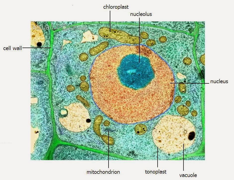
A LEVEL SCIENCE NOTES: Plant cell structure
